Magnetic resonance
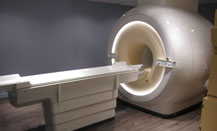
This technique uses a powerful magnetic field and radiofrequency waves. No ionizing radiation is used. MRI is most useful to diagnose lesions that cannot be imaged with standard radiographs, ultrasound or computed tomography.
The maximum weight for this exam is 350 pounds. We are sorry for any inconvenience.
Radiologie Varad is proud to offer Gadovist, a macrocyclic and non-linear product to our clients for examinations with injection, therefore without depositing Gadolinium in the brain.
- MRI Abdomen
- MRI Circle of Willis
- MRI Cranio-vertebral junction
- MRI Neck - ENT
- MRI Breast
- MRI Musculoskeletal
- MRI Carotid
- MRI Brain
- MRI Internal auditory canals
- MRI Pituitary gland
- MRI Orbits
- MRI Pelvis
Contact us now by calling at 514 281-1355
Request an appointment Questions? Contact usComputed Tomography
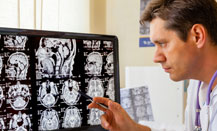
The scan uses X-rays that are emitted in a circular pattern around the patient and captured by detectors located on the opposite site of the emitter. From this information, the computer reconstructs images that represent the transverse slices of the body. The new machines are very fast and an exam can be performed in less than five minutes.
- Virtual Colonoscopy
- Scan Abdomen
- Scan Brain
- Scan Facial bones
- Scan Orbits
- Scan Pelvis
- Scan Sinuses
- Scan Coronary Calcium Scoring
- Scan SI Joints
- Scan Neck - ENT
- Scan Mastoids – Temporal Bones
- Scan Musculoskeletal
- Scan Lumbar spine
- Scan Thorax
Contact us now by calling at 514 281-1355
Request an appointment Questions? Contact usUltrasound and Doppler
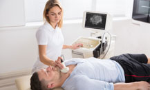
Ultrasound is a simple non-invasive examination, using an ultrasound beam to create an image by moving a probe on the skin surface. The Doppler technique allows for a more specific examination of blood vessels.
Ultrasound is used for abdominal and pelvic exploration, more specifically for the liver, kidneys, aorta, pancreas, gall bladder, uterus and ovaries. It is the preferred examination to monitor pregnancies. Ultrasound tests are also used to assess muscles, tendons, and other peri-articular structures.
Doppler exam evaluates blood flow; it can detect narrowing and occlusions of blood vessels. It is used for the carotid and vertebral arteries, the intracranial vessels, and the arteries and veins of the abdomen and extremities.
- Doppler Venous (Trombophlebitis only)
- Ultrasound Abdomen
- Ultrasound Breast
- Ultrasound Fetal (16 weeks or less)
- Ultrasound Nuchal translucency
- Ultrasound Pelvic (+/- endovaginal)
- Ultrasound Testicles
- Ultrasound Thyroid
- Ultrasound Musculoskeletal **New April 2022**
Contact us now by calling at 514 281-1355
Request an appointment Questions? Contact usMammography

Mammography is a specialized examination of the breasts for the early detection of breast cancer and diseases of the breast. The exam is done with the use of specialized equipment especially designed for the breast.
This compression is needed for the following reasons:
- It decreases the amount of radiation needed by decreasing the thickness of the breast;
- It limits movement during the exposure;
- It separates the breast tissue to allow better evaluation of lesions that could be hidden by the breast tissue;
- It creates homogeneous thickness of the entire breast allowing a more uniform penetration of the tissue;
- It increases the precision and detail of the breast by keeping the tissue closer to the film.
Two types of mammography:
- Diagnostic Mammography
- Screening Mammography
Screening Mammography
Our mammographic technician takes all necessary precautions to minimize the pain that you may feel. If you did feel any pain during the compression, there is no cause for concern. This pain is temporary and without consequence.
Mammography is the most efficacious study to detect small breast cancers, but it does not detect all cancers. It is therefore important to practice regular breast self-examination. Post-menstrual is the best moment to identify breast changes. It is not easy to examine a breast but if you do it on a regular basis every month, after a year, you will have performed twelve examinations and you will know the feeling of your breasts and can then report any unusual changes there may be.
Our clinic is appointed by the Québec Breast Cancer Screening Program.
New generation Philips MicroDose mammograph, guaranteeing a rapid examination, with a lower radiation exposure and a great quality image.
Contact us now by calling at 514 281-1355
Request an appointment Questions? Contact usMusculoskeletal Imaging
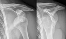
Consultation, imaging and interventional procedures in musculoskeletal pathologies such as:
- Tendinopathy
- Bursopathy
- Retractile capsulitis of the shoulder
- Calcifying tendinopathy of the shoulder or impingement syndrome of the shoulder
Our team of radiologists from the CHUM, specialized in musculoskeletal imaging and interventional radiology, will insure specialised treatments, in a comfortable environment, all the while favouring communication and a multidisciplinary approach with the referring physician.
- Radiography
- Scan (computed tomography)
- Arthro-scan
- Magnetic resonance imaging (MRI)
- Arthro-resonance imaging
- Ultrasound and ultrasound guided therapeutic injections
- Therapeutic injections under fluoroscopy
- Corticosteroids
- Visco-suppleance (Synvisc, Durolane, etc)
Contact us now by calling at 514 281-1355
Request an appointment Questions? Contact usGeneral radiology
Despite the highly sophisticated technical developments of computed tomography scans, ultrasound and magnetic resonance imaging, conventional radiographs remain an essential diagnostic tool.
Osteo-articular
- Cervical Spine
- Dorsal Spine
- Lumbar Spine
- Sacrum-coccyx
- Limbs
- Sacro-iliac joints
- Pelvis
- Hip
- Lower extremities (lying position)
- Scoliotic series 18 years old and over
- Bone series
- Articular series
Head and neck
- Skull
- Facial Bones
- Temporomandibular joints
- Cavum
- Lower Jaw
Chess-Thorax
- Chess
- Thorax
Abdomen
- Simple cliché
- Abdominal series
Fluoroscopic study
- Hysterosalpingography
- Arthrography
- Bursography
- Facet block
- Therapeutic infiltrations
Contact us now by calling at 514 281-1355
Request an appointment Questions? Contact usOsteodensitometry

As you know, osteoporosis has become a more common medical problem with the aging population. Clinical research in this area is very active and has brought new therapeutic alternatives.
Over the recent years, there have been many new techniques to measure the bone density. Amoung the most commonly used exams; osteodensitometry is the study of choice. Its precision is superior to all other methods and allows easy follow-up of patients. The exposure to ionizing radiation is minimal and inferior to other methods. The exam is short in duration, around 5 minutes, and is comfortable for the patient.
The aims of the study are to:
- Establish the indications for preventative hormone replacement at menopause;
- Determine the bone density in patients who are at risk of developing severe osteoporosis;
- Establish the diagnosis of osteoporosis when the radiograph raises a suspicion;
- Follow the treatment response against osteoporosis.
Contact us now by calling at 514 281-1355
Request an appointment Questions? Contact us